A Hen Laid a Matreshka
Newspaper Trud, Moscow, September 26, 1981
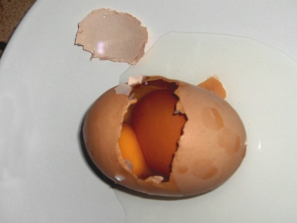
Translated from Russian by Michael RomanovThe examiner of the M. Gadzhiev Makhachkala Plant, L. Arteiva, has a small subsidiary plot. And one day one of her hens laid an extraordinary egg, round and goose egg-sized. When it was broken, one more, smaller, egg occurred inside.
This is a rather seldom phenomenon, the newspaper Dagestanskaya Pravda says, as Dr. N. Grigoryeva, Candidate of Agricultural Sciences (Ph.D.) and Assistant Professor of the Daghestan Agricultural Institute, explains. The common people call such eggs matreshkas [usually wooden Russian doll in peasant dress with successively smaller ones fitted into it].
An egg contained another egg was laid by a hen in the small French town Baneuil, its whole weight being 170 g. But the champion was perhaps a hen from Yugoslavian village Zagunjica, she laid an egg weighing more than 200 g and having three yolks.
Comments
by Professor Randall K. Cole
Unit of Avian Medicine, Department of Microbiology and Immunology
College of Veterinary Medicine
Cornell University, Ithaca, N.Y. 14853-6401, USAFrom his letter of December 15, 1998, to Michael Romanov:
The reference to Matreshka brings back to memory of double yolk eggs found here [at Cornell University] by Professors Alexis Romanoff and Frederick Hutt back in the 1940's (Romanoff and Hutt, 1945). Hutt discusses such variations in eggs, in his book - "Genetics of the Fowl" (Hutt, 1949, pp. 393-398). He cites one that weighed 227 g and contained a shelled egg plus two additional free yolks. It came from a Rhode Island Red in Scotland (Brown, 1939).
It also reminds me of a retirement party for Professor Graham, who was head of the Poultry Department at the Massachusetts College, in Amherst. He was sent a nice egg from the poultry staff at the Kansas State College, with instructions for his wife to hard-boil it and bring it to the party where she would receive further instructions. There she had to break the shell and hand the contents to Professor Graham. The albumen was cooked, but when he separated it from the "yolk", he found a unit not quite like a normal yolk. It turned out to be a letter of congratulations, with signatures from the staff at Kansas. [Professor Peter Sharp, Division of Development and Reproduction, Roslin Institute (Edinburgh), Scotland, considers this trick possible by means of surgery and transplantation of a capsule into the hen's oviduct (Sharp, personal communication, 1998)]
Here on my desk I have a photograph of Prof. Hutt, taken at his retirement in 1965 which brings back many memories. He earned his Ph.D. degree at the University of Edinburgh (1929). Later he was honoured there with the D.Sc. degree in Genetics (1939). After retirement, he was made an Honorary Fellow of the Royal Society of Edinburgh.
References
Brown, J.M. (1939) Scotland claims a world egg record. Farmer and Stock-Breeder, 53: 746.
Hutt, F.B. (1949) Genetics of the Fowl (New York, McGraw-Hill Book Company, Inc.).
Romanoff, A.L. and Hutt, F.B. (1945) New data on the origin of double avian eggs. Anatomical Record, 91: 143-154.
Ovum in ovo
by Francis H. Herrick
The American Naturalist, vol.33, No.389, May 1899
I.
An interesting case of egg within egg recently came to hand, which was unique in this respect: that the smaller enclosed egg lay in the yolk and not in the albumen of the surrounding egg, as in all similar instances hitherto recorded. This hen’s egg had unfortunately been cooked. When it was opened, and the white broken into, a suspicious-looking fleck was seen on the surface of the hardened yolk, the removal of which disclosed the imbedded egg. The parts of the containing egg have not been preserved, so that I can record only the general facts here given.
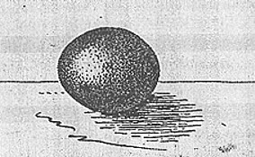
The included egg measures 17 x 21 mm., is of symmetrical ovoidal form, possesses a hard shell, shell membrane, and small yolk. The shell has a coarse granular texture, and a coffee color, sprayed with brown pigment. Its form and appearance are represented in Fig. 1, its relation to the containing egg in Fig. 2.
Fig. 1. Small egg removed from yolk of larger egg.
Natural size.Since writing the foregoing and following parts of this paper, I received a second example of ovum in ovo, which is represented in Fig. 3. The smaller enclosed egg lies in the albumen of the larger, as in the cases hitherto described. It measures about 18 x 22 mm., has a clear shell of even texture, shell membrane, and albumen. Apparently no yolk is present; at least none could be found in the specimen, which has been somewhat mutilated. The parts of the surrounding egg appeared normal in every respect.
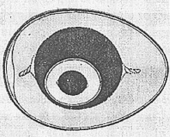
Fig. 2. Diagram to show the relations of ovum in ovo, in which the enclosed egg (Fig. 1) lay in the yolk. This represents the relative form and size of the included egg, and shows its general relation to the yolk of the enclosing egg. In other respects the figure is conventional. Shell, shell membrane, albumen, and yolk are represented in each egg. About four-fifths natural size.
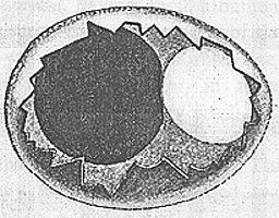
Fig.3. Diagram to show the relations of ovum in ovo, in which the included egg lay in the albumen. The shell is represented as broken, exposing the yolk shaded dark, and to the right the small enclosed egg, which is white. About four-fifths natural size.
II.
These cases belong to that class of abnormalities in the eggs of birds which originate before incubation, and to the variety called ovum in ovo. In all recorded cases of this type the contained bodies lie in the albumen, or at least not in the yolk of the surrounding egg.
In 1878 Parona and Grassi (Sovra alcune monstruositŕ di uova di gallina, Atti della Soc. Ital. di Sci. Nat., Milano, vol. xx (1878)) described and figured a good example of the more common condition in which the in egg, an elongated ovoidal body, lay in the albumen. The yolk of the larger egg possessed a normal blastoderm. These’ gave a history of monstrosities of this class known up to that time, from which the following abstracts are drawn.
Davaine, in a work published in 1860, gave a résumé of cases of ovum in ovo then known. He found that the enclosing egg might be larger or smaller than normal, having shell, albumen, and yolk, the latter liable to deformity from disturbance of the foreign body. The included egg was very rarely of normal size; usually it was small and devoid of yolk. Clayer, in 1682, described a case where the included egg was very small, but possessed a yolk.
A similar example was also reported by Yung in 1671. The yolk of the inside egg was very small, and possessed two chalazae. Rayer described, in 1849, a goose egg of colossal size, which contained an egg of normal dimensions, having yolk, albumen, and shell. The outer egg was also complete, although its yolk was flattened by pressure. A few years later three similar cases were reported by De Moroga, Aucapitaine, and Alessandrini. The latter reported the case of an egg. of normal size, which contained a small egg complete in all but the shell.
In 1856 a case is cited, by Davaine, of three eggs enclosed by a common shell, and still later a precisely similar phenomenon was described by Flourens.
Panum, who studied for several years the abnormalities of birds’ eggs and collected many examples, met with the ovum in ovo only once. This resembled Rayer’s goose egg, and came from the Indian jungle fowl. The inside body corresponded to the normal egg in size, while the surrounding egg was of relatively huge proportions. Panum adds that in 1858 a remarkable egg of a Cochin China hen was described. It was very heavy and contained two yolks and one ordinary egg with solid shell. Still another egg, analogous to that of Rayer’s goose, was described by Bert in 1861. Parona and Grassi remark that the foregoing are the only cases recorded of this singular abnormal production, but that it would be possible to cite more than thirty instances in which small yolkless eggs have been found enclosed in another egg apparently normal. Later additions to this literature have been made by Landois in 1882, and by Schumacher in 1896.
Landois remarks that in most cases of ovum in ovo the included egg is small and yolkless, and sometimes possesses a very abnormal form. In one erratic individual it resembled a tapeworm, consisting of a button the size of a pin head, followed by a fibrous section, and ending in a broad flattened string. He thinks that most so-called tapeworms in hens’ eggs are nothing more than monstrosities of this kind.
In the case described by Schumacher the enclosing egg was apparently normal, and, like that figured by Parona and Grassi, possessed an egg-like inclusion in the albumen. This was of regular oval form, a little larger than that here described, and, besides hard shell, possessed a stratified albumen, an irregular spirally twisted yolk mass, measuring 8 x 4 mm., with small chalazae at either end. There was no blastoderm.
III.
Reviewing the cases of ovum in ovo in zoological literature, we may classify them in the following manner:
I. Enveloping egg usually normal, but occasionally of large size; blastoderm recorded in at least one instance.
Enveloped egg:
(a) In yolk; small; composed of shell, shell membrane, albumen, and yolk; no blastoderm known to occur in this or in the following variations; single case recorded in this paper (Fig. 2).
(b) In albumen; small; composed usually of shell, shell membrane, albumen, and rarely with yolk; few cases reported.
(c) In albumen; small; usually with shell, shell membrane, and albumen, but no yolk; most cases of ovum in ovo reported are of this kind (Fig. 3).
(d) In albumen; usually small and variously distorted, so as to bear little resemblance to an egg at all.
II. Enveloping egg of colossal size; complete; blastoderm probably present.
Enveloped egg:
(a) Without shell, but otherwise complete. In this case a common shell may surround two or three eggs complete except for shells and shell membranes; forming "double-yolk" or "triple-yolk" eggs.
(b) One of the enclosed eggs of normal appearance and size, possessing shell, albumen, and yolk; the other eggs surrounded by a common shell, but having no shells of their own.
According to the accepted accounts, the yolk of the fowl’s ovum is of normal size when it leaves its vascular capsule in the ovary, and is taken up by the infundibulum. Reaching the oviduct it receives its first layer ‘of albumen, and is carried slowly down the tube by peristaltic muscular contraction of the walls. Near the lower end of the oviduct the final layers of albumen are added, and in the distal extremity of the duct, called the uterus, the shell is formed.
In the opinion of several writers the small included egg represents a fragment of a normal ovum which has been ruptured, and thus has parted with some of its substance after leaving the ovary. This fragment is then treated in the oviduct like a full-sized egg. The small egg-like body thus produced is sometimes laid, but occasionally it is driven by anti-peristaltic action up the tube until it collides and fuses with the mother egg. This theory will suffice to explain the first class of inclusions on the supposition that rupture takes place in .the upper part of the oviduct, or at least after the first layers of albumen have been added to the normal egg. Since the small included eggs are generally yolkless, we must infer that such ruptures are, as a rule, confined to the albumen. It seems more probable that the small egg becomes enclosed before the shell membrane is formed over a normal egg, with little if any retrogressive movement. Furthermore, it is possible that any substance which serves as a local stimulus to the upper part of the oviduct, whether coining from the ovary as abortive egg or egg-fragment, or from the duct as secreted product, may serve as a nucleus, about which an egg-like body may be formed. Normally laid eggs, indeed, tend to sweep the oviducal canal clear of all obstructions, whether they be abortive eggs, blood clots, feathers which rarely grow from its walls, or parasites which find their way into it. These bodies are taken up by the egg, and become imbedded in it. In one of the cases recorded in this paper the obstruction or small "egg" (Fig. 2) has been pressed through the albumen, through the vitelline membrane, and thus into the yolk itself. It is evident that inclusions of the first type are not true eggs in any strict sense, since, so far as known, they contain no protoplasm.
The theory of yolk-hernia will not explain the second class of abnormalities, such as double or triple yolk eggs. We have here a case of fusion of the albumen in two or more ova, which are treated in the uterus as one egg and surrounded by a single shell. This process is sometimes complicated by the inclusion of a third egg of normal size and already covered by a hard shell. These conditions may be brought about by irregularities in the mechanism of the oviduct, as when any given egg does not receive its shell and is not laid before it encounters others coming down the oviduct at the same time.
According to Duval, an egg usually spends thirty hours in the oviduct, twenty-four of which are passed in the uterus, and if the fowl lays once in forty-four hours a single egg will be found in the oviduct at any given time. When the intervals of laying become shorter, however, an egg may be found at either end of the tube. In some of the cases described three ova must have been in the tube and collided there, owing to disturbances in the normal rhythms.